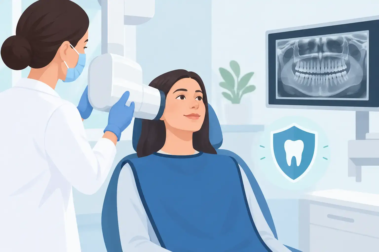
A cavity can begin between two teeth, beneath an old filling, or below the gumline – places even a careful visual exam cannot fully show. That is why patients often ask, are dental x rays safe? It is a reasonable question, particularly if you are pregnant, bringing in a child, have had several medical scans, or simply prefer to limit radiation whenever possible.
For most patients, dental X-rays are considered safe when they are recommended for a clear clinical reason and performed with appropriate safeguards. The radiation dose from a typical dental image is low, but low does not mean unnecessary. A thoughtful dentist weighs the value of the information against your health history, symptoms, past images, and risk for dental disease.
Are Dental X Rays Safe for Routine Dental Care?
Dental X-rays use ionizing radiation, the same broad type of energy used in some medical imaging. Ionizing radiation can affect living tissue at high enough doses, which is why dental teams follow the principle of ALARA: “as low as reasonably achievable.” In plain terms, they use the least radiation needed to obtain an image that can guide care.
According to the American Dental Association and the U.S. Food and Drug Administration, dental radiographs are a safe and useful diagnostic tool when selected based on an individual patient’s needs. Modern digital imaging generally requires less radiation than older film-based methods, although the exact dose varies by the type of image, equipment, settings, and technique.
The risk is not viewed in isolation. We all receive small amounts of background radiation from natural sources in daily life, including the ground, air, and food. Dental X-rays add a small amount to that background exposure. Still, comparing doses should not replace a personal conversation about whether a particular image is needed for you.
Why a dentist may recommend an X-ray
An X-ray can reveal concerns that are not visible during a regular exam. These may include decay between teeth, infection around a tooth root, bone changes related to gum disease, impacted teeth, cysts, or problems beneath an existing restoration. Images can also help a dentist plan certain treatments and monitor changes over time.
Finding a concern early may allow for a simpler approach. For example, decay detected while it is small may be easier to address than decay that has progressed toward the nerve. That does not mean every visit requires new X-rays. It means images should be taken when they are likely to change the dentist’s understanding or treatment recommendation.
How Often Should Dental X-Rays Be Taken?
There is no single schedule that fits every adult or child. A person with healthy teeth, stable gums, and a low risk of cavities may not need bitewing X-rays as often as someone who has frequent decay, dry mouth, many restorations, active gum disease, or new dental symptoms.
Your age and dental history matter, too. Children and teens may need images at different intervals because their teeth and jaws are developing and they can be more prone to cavities. A patient who is new to a practice may need diagnostic images if prior records are unavailable or do not provide the information needed for a complete exam.
A good question to ask is: “What are you looking for with this X-ray, and do you have recent images I can transfer?” If you had X-rays taken at another dental office, tell the team. Existing images may be useful, depending on their quality, age, and the reason for today’s visit.
Different Dental X-Rays Have Different Purposes
Not all dental images expose the same area or answer the same question. Bitewing X-rays typically show the crowns of back teeth and are commonly used to look for decay between teeth and changes in the bone supporting the teeth. Periapical X-rays show an entire tooth, including the root and surrounding bone, and can help investigate pain, infection, or a localized concern.
A panoramic X-ray captures a broad view of the jaws, teeth, and surrounding structures. It may be helpful when evaluating impacted teeth, jaw concerns, or general development. Three-dimensional imaging, often called cone beam CT or CBCT, can provide more detailed information for selected situations, but it typically involves more radiation than standard dental radiographs. It should be used when the added detail is likely to affect care.
The key point is that a larger or more detailed image is not automatically “better.” The best image is the one that answers the clinical question with the appropriate level of exposure.
What About Dental X-Rays During Pregnancy?
If you are pregnant or think you might be pregnant, tell your dental team before any imaging is taken. Dental care should not automatically be put on hold during pregnancy, especially when there is pain, swelling, suspected infection, or another urgent concern. Untreated dental infection can create its own health issues.
The American College of Obstetricians and Gynecologists states that dental X-rays with appropriate shielding are considered safe during pregnancy when clinically necessary. Your dentist can discuss why an image is recommended now, whether it can reasonably wait, and what precautions will be used. Routine imaging that is not needed right away may be postponed, while diagnostic imaging for an active problem may be appropriate.
This is also a time to share any concerns openly. You deserve an explanation that feels clear, not rushed.
How Dental Teams Reduce Exposure
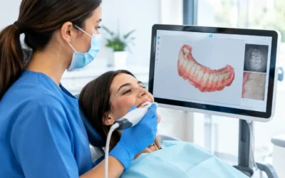
Radiation safety is more than placing a protective apron. It starts with deciding whether an image is necessary at all. Dental teams can reduce exposure by using digital sensors when available, selecting the right image type, limiting the number of images, using proper positioning, and avoiding repeat images caused by movement or technique issues.
Protective shielding may be used based on the practice’s protocols, the patient’s needs, and current guidance. Recommendations on shielding have evolved as equipment and safety standards have improved. If you have questions about a thyroid collar, lead apron, or another precaution, ask before the image is taken. The team should be able to explain its approach in straightforward language.
You can also help by holding still, following positioning instructions, and letting the team know if you have trouble tolerating a sensor. Discomfort from a sensor is common for some people, but adjustments may be possible.
When It Makes Sense to Ask More Questions
It is appropriate to pause and ask for more context, especially if you are unsure why imaging is being recommended. This is not being difficult. It is participating in your care.
Consider asking how the image will affect the examination or treatment plan, whether recent images can be used instead, what type of X-ray is proposed, and what alternatives exist. In some cases, there may be no equally useful alternative. In others, monitoring a concern or using a different image may be reasonable. The answer depends on your symptoms, oral health history, and the information already available.
If you have had radiation treatment, have a complex medical history, or are concerned about cumulative exposure from other imaging, mention it. Your dentist can consider that information while coordinating with your medical providers when appropriate.
A Calm, Informed Conversation Helps
Dental X-rays are not something to fear or accept without explanation. They are one diagnostic tool, used to help a dentist see what the eye cannot. When images are chosen carefully and taken with current safety practices, their benefits often outweigh the very small radiation risk.
At West Hollywood Smile Dental, patients can ask why an image is being recommended, what it may show, and how it may shape their care options. Whether you are due for a checkup, dealing with a new concern, or preparing for restorative or cosmetic treatment, a consultation is a good opportunity to discuss imaging at a pace that feels comfortable and informed.